NANOSCOPIUM is a hard X-ray nanoprobe beamline, which is dedicated to fast scanning multi-modal and multi-lengthscale imaging. It offers simultaneous information in a quantitative manner and in the same experimental conditions, in 2D & 3D about the elemental composition, chemical speciation and sample morphology. Namely, Nanoscopium offers a large portfolio of complementary nano-imaging and spectro-microscopy methods, also in association with full field micro-tomography techniques. The cutting edge multi-modal possibilities are available at the beamline at hierarchical length-scales in order to tailor, through several orders of magnitude, the field of view (µm-mm) and spatial resolution (70 nm – 1µm) to the experimental needs.
Available analytical techniques:
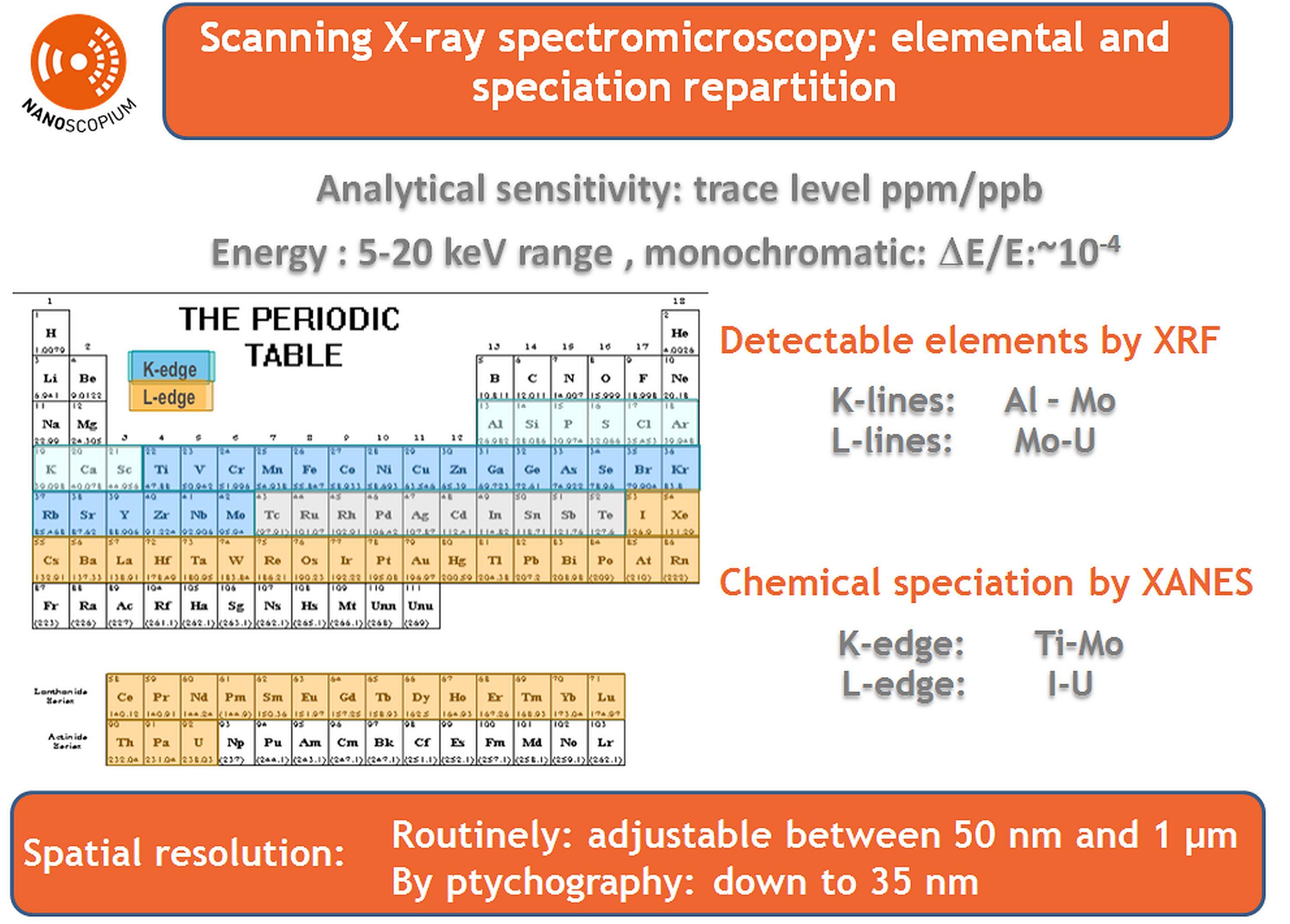
- X-Ray Fluorescence (XRF) nanoprobe: open for user applications
- X-ray Absorption Spectroscopy (XANES): open for user applications
- Full field X-ray microtomography: open for user applications
- XRF tomography: please contact the beamline scientists
- Scanning X-ray Diffraction: please contact the beamline scientists
Acquisition modes:
- 2D at multiple lengthscales
- 3D by full field microtomography tomography & scanning tomography
- Fast continuous sample scanning (FLYSCAN, multi-detector architecture) with down to deca-ms dwell time/pixel
- Network architecture: 10 Gbits tailored to the au high data flux produced by the ensemble of detectors (1 TOctets per day)
The nanoprobe stations in exploitation in sequential mode:CX2 station:
Full field X-ray microtomography in absorption and phase contrast modes
KB-based nanoprobe, CX3 :
- Nanofocusing optics: chromatic, KB (JTEC)
- Fast multimodal and multi-lengthscale imaging with spatial resolution down to 70 nm. High photon flux providing high analytical sensitivity (ppm range) for elemental and chemical speciation characterization
Cryo-U18
Fixed exit Double Crystal Si(111)
CX2 station
CX3 station
- Nanofocusing optics: achromatic diffraction limited focusing, High optical quality Kirkpatrick-Baez (KB) mirror-pair (JTEC), its opto-mechanical system has been provided by Bruker.
Entrance Optics
Fresnel-Zone based nanoprobe, CX2
Sample
KB-based nanoprobe, CX3
Fast multimodal and multi-lengthscale imaging for elemental (scanning XRF), chemical characterisation (XANES, spectromicroscopy) and morphological (scanning differential phase contrast and dark field) characterisation.
Spatial resolution at sample: ~70 x 70 nm2 to 1 x 1 µm2 by FLYSCAN.
Flux on sample: 10^10 ph/s at 15 keV
Available detectors:
Single element Si Drift Detector (SDD, Ketek)
Fast digital multichannel analyser:
4-channel XMAP (Xia, inc)
Sample
Pixel-detector: MerlinX
Detection
Single element Si Drift Detector (SDD, Ketek)
4-channel XMAP (Xia, inc)
Detection
X-Ray camera with inderect conversion: Scintillateur + optics with magnification of (G4) PCO.Edge
Detection
X-Ray camera with inderect conversion: Scintillateurs + optics with magnifications of (G2, G5, G10) ORCA FLASH
Detection
- X-ray fluorescence (XRF)
- X-ray microscopy
- X-ray tomography
- Biochemistry
- Geology
- Global change & Climate observation
- Marine science/Oceanography
- Natural disaster, Desertification & Pollution
- Other - Earth Sciences & Environment
- Plant science
- Arts
- Cultural Heritage
- Biophysics
- Other - Life Sciences & Biotech
- Astronomy/Astrophysics/Astroparticles
- Tango, Spyc
- elemental distribution maps, morphology distribution maps, XANES spectra, absorption and phase contrast microtomograms
- nxs/hdf5 format
- PyMCA, ImageJ